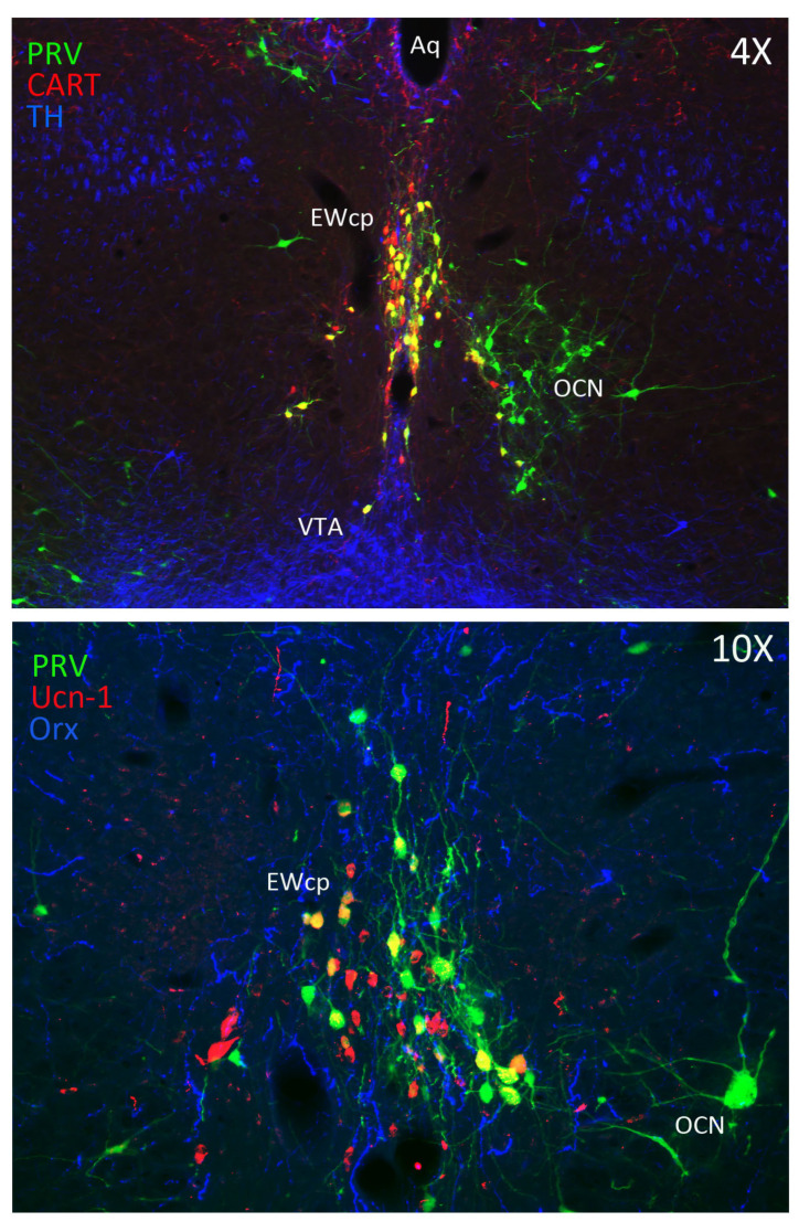
Figure 1.
Infected neurons in the EWcp after PRV injection into BAT in rat. Brain sections were processed for triple immunofluorescence. Upper image shows infected neurons labeled in green, CART-ir neurons in red, and dopaminergic neurons (tyrosine hydroxylase-ir) in blue at low magnification (4×). Most infected neurons in EWcp are CART-ir (double-labeled in yellow), intermingled with some non-infected dopaminergic neurons (blue). Lower image shows infected neurons labeled in green, Ucn-1-ir neurons in red and Orx-ir fibers in blue at high magnification (10×). Dense Orx fibers appear in close contact with infected Ucn-1-ir neurons, which are double-labeled (yellow). Bregma level = −6.00 mm (upper image) and −5.62 mm (lower image) based on the rat brain atlas from Paxinos and Watson, 6th ed. [13]. Abbreviations: EWcp, Edinger-Westphal nucleus; Aq, aqueduct; OCN, oculomotor nucleus; TH, tyrosine hydroxylase; VTA, ventral tegmental area.

