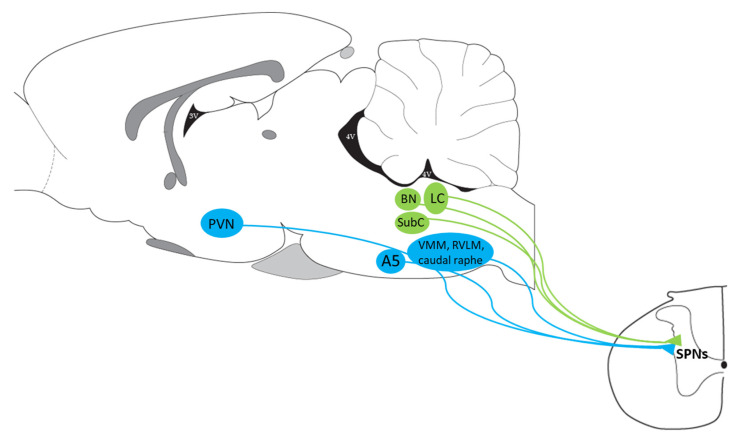
Figure 2.
Presympathetic brain regions that project directly to the IML of the spinal cord where SPNs are located. Classical presympathetic areas are depicted in blue, whereas those in green are the regions we suggested to be added based on PRV neuronal tracing. For terms, see the list of abbreviations.

