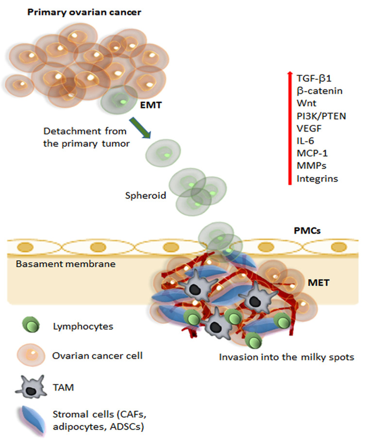
Figure 1.
Schematic representation of metastatic pathways of ovarian cancer. The initial steps of metastasis required several mechanisms, including suppression of adaptive immune response, ECM reorganization, and the promotion of epithelial-mesenchymal transition (EMT). During initial tumorigenesis, ovarian carcinoma cells undergo EMT, which involves a change in integrin expression and up-regulation of different signaling pathways (Wnt, β-catenin, PI3K, IL-6), angiogenic (VEGF), and proteolytic molecules (MMPs). Cancer cells can detach from the tumor and, carried by the peritoneal fluid, cancer cell spheroids overcome anoikis and attach preferentially on the abdominal peritoneum. Cell aggregates find preferential engraftment into the omental “milky” spots, composed of lymphocytes, macrophages (TAMs), and stromal cells (CAFs, adipocytes, and ADSCs). At the peritoneal interface, cancer cells invade peritoneal mesothelial cells (PMCs) facilitated by integrins and TGFβ1, released by tumor cells and CAFs. Ovarian cancer cells undergo MET (reverse EMT) to acquire an epithelial phenotype, enabling the cells to establish and grow, developing secondary tumors and metastasis. Abbreviations: ECM, extracellular matrix; EMT, epithelial-mesenchymal transition; VEGF, vascular endothelial growth factor; MMPs, matrix metalloproteinases; TAMs, tumor-associated macrophages; CAFs, cancer-associated fibroblasts; ADSCs, adipose-derived stem/stromal cells; PMCs, peritoneal mesothelial cells; TGFβ, transforming growth factor-β; MET, mesenchymal-epithelial transition.

