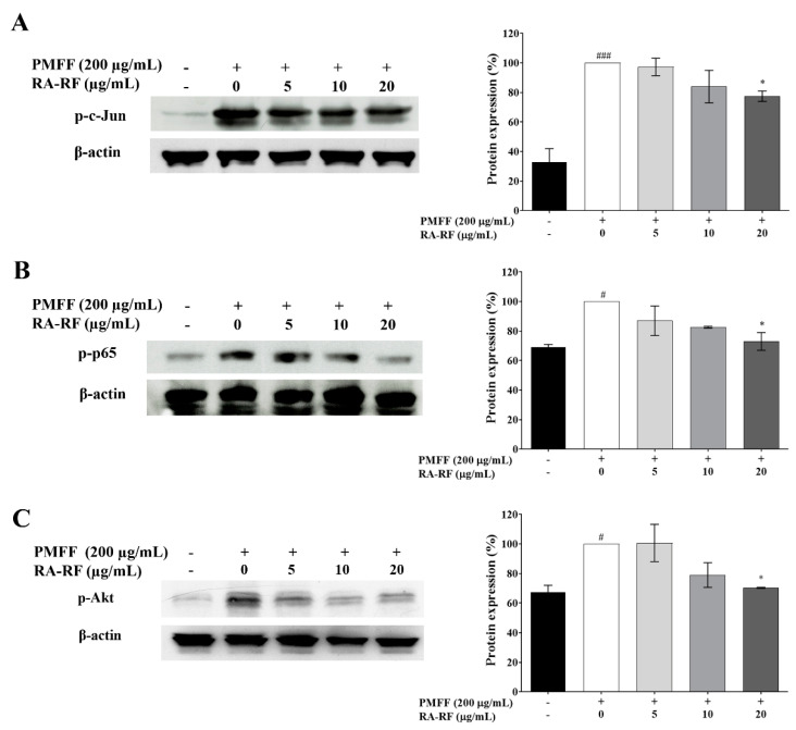
Figure 5.
The protein expression of p-c-Jun (A), p-65-NF-κB (B), and p-Akt (C) in A549 cells exposed to PMFF (200 µg/mL) in the presence of RA-RF (5, 10, and 20 µg/mL) determined by Western blot analysis. Error bars indicate SD. The mean ± SEM are shown as # p <0.05, ### p < 0.001 vs. the control group; * p < 0.05 vs. PMFF group. The data were independently performed in triplicate.

