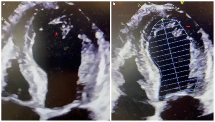
Figure 3. Patient's transthoracic echocardiogram.
(a) Apical four-chamber view demonstrating the severe apical akinesis with left ventricular dilatation as depicted by the red star (apical ballooning). (b) Apical four-chamber view with endocardial tracing outline illustrating the similarity to the Japanese octopus trap as depicted by the red arrow.

