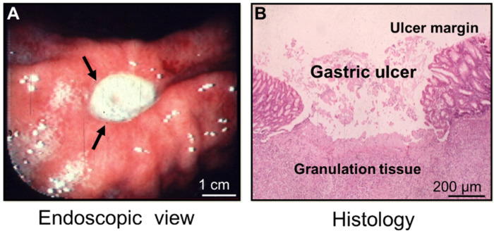
Figure 1.
A gastric ulcer—endoscopic view and histology (reprinted with permission from [1]). (A) An endoscopic picture of a human gastric ulcer on the lesser curvature of the stomach (arrows). The ulcer is covered with white fibrinopurulent material. (B) A histological image of an experimental gastric ulcer in a rat model; hematoxylin and eosin (H&E) staining were used.

