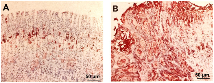
Figure 4.
Photomicrographs of rat gastric oxyntic mucosa immunostained for epidermal growth factor receptor and counterstained with hematoxylin. (A) Normal rat gastric oxyntic mucosa shows epidermal growth factor receptor localization only in some neck cells of gastric glands (the progenitor zone; brown staining). (B) Mucosa at the GU margin 7 days after ulcer induction shows the “disappearance” of parietal and chief cells due to their dedifferentiation and reprogramming to progenitor cells (brown staining) through increased expression of epidermal growth factor receptors in the majority of cells lining GU margin. The cells demonstrating epidermal growth factor receptor expression occupy the entire mucosal thickness (reprinted with permission from [15]).

