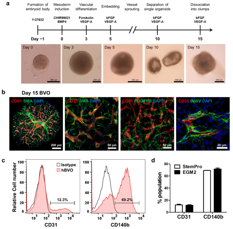
Figure 1.
Generation of blood vessel organoids. (a) Schematic diagram of the protocol. (b) Endothelial networks showing the establishment of blood vessel organoids and vascular networks coated with pericytes (PDGFRβ+), smooth muscle cells (SMA+), and basement membrane (collagen type IV [ColIV]+). (c), Representative FACS histogram showing the percentages of endothelial cells (CD31+) and pericytes (CD140b+) in the blood vessel organoids (n = 9). (d) Histogram showing no significant differences in the cell population (endothelial cells and pericytes) of hBVOs between culture media (StemPro-34 and EGM2). Bars and error bars represent the mean ± SD of results in triplicate experiments (n = 9); unpaired, two-tailed student’s t-test.

