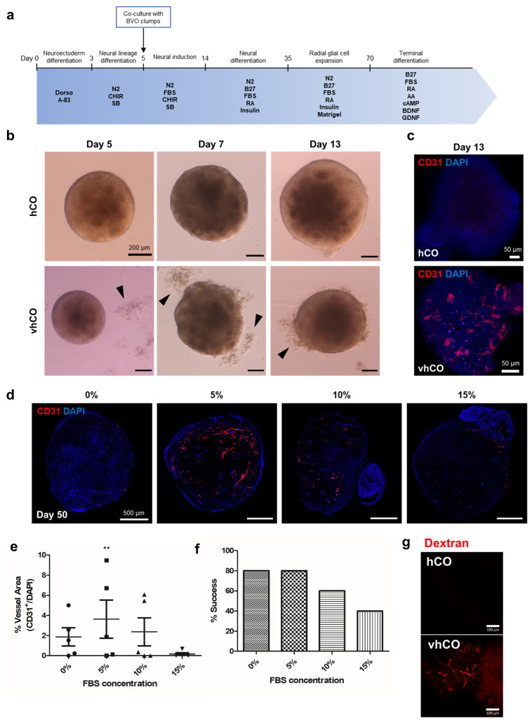
Figure 2.
Generation of vascularized cerebral organoids. (a) Schematic diagram of the procedure used to generate vascularized cerebral organoids. (b) Blood vessel clumps (black arrowheads) fused to neural organoids generated on day 5 in the co-culture of developing cerebral organoids. (c) CD31+ endothelial cells were observed on day 13 of vhCO development. (d) CD31+ endothelial networks generated under different FBS conditions. (f) The percentage of organoids succeed to generate vessels after co-culture in different FBS concentrations. (e) Quantification of vessels performed using AngioTool. Data are plotted as means ± standard deviation. ** p < 0.01; one-way ANOVA. (g) Dextran was perfused on vhCO. (f) Ratios of successful vascularization under various FBS concentrations.

