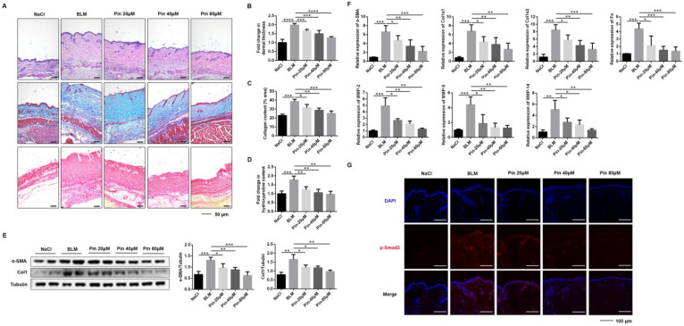
Figure 5.
Pinocembrin ameliorates BLM-induced skin fibrosis in mice. (A) Representative skin sections stained with hematoxylin–eosin (H&E), Masson’s trichrome, and Sirius red staining (×100, Scale bar = 50 μm). (B) Total dermal thickness of the back of each group of mice. (C) Collagen density was quantified on Masson’s trichrome images. (D) Hydroxyproline content of skin tissues in mice. (E) Protein levels of α-SMA and Col1 were verified by Western blot in the lesional skin. (F) mRNA levels of α-SMA, Col1α1, Col1α2, Fn, MMP-2, MMP-9, and MMP-14 in the lesional skin were assessed by qPCR. (G) Immunofluorescence staining of p-Smad3 in skin frozen sections of BLM-induced model (×400, Scale bar = 100 μm). Means± SD, n = 8. * p < 0.05, ** p < 0.01, *** p < 0.001, **** p < 0.0001.

