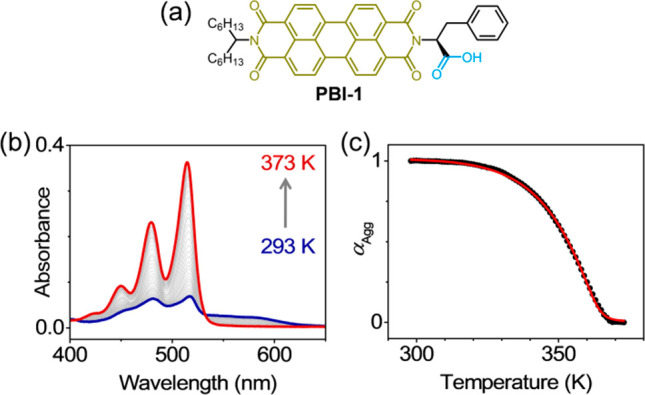
Figure 1.

Cooperative self-assembly of PBI-1. (a) Molecular structure of PBI-1, showing the motifs responsible for π-stacking (olive) and hydrogen-bonding (blue). (b) Temperature-dependent optical absorption spectra of PBI-1 aggregates in methylcyclohexane (15 μM). (c) Variation of αAgg with temperature, derived from the 0–0 absorbance at 515 nm. The red line shows the simulated curve generated using mass-balance model for σ = 6.3 × 10⁻⁴, at 293 K.
