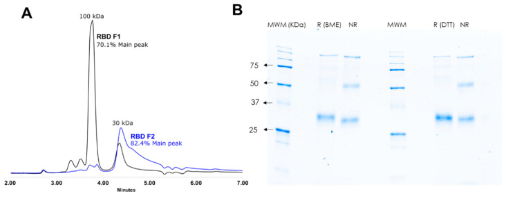
Figure 1.
Characterization of the RBD protein. The RBD protein was expressed in HEK 293 cell cultures and purified on a HisTrap™ Nickel column; two fractions were obtained. (A) Native SEC analysis evinced that Fraction 1 (RBD F1; black line) was mainly composed of a ~100 kDa protein and Fraction 2 (RBD F2; blue line) by a ~30 kDa protein, which may correspond to the trimer and monomer of the RBD protein, respectively. (B) Fraction 2 also exhibits the main band at ~30 kDa when analyzed in denaturing SDS-PAGE using two reducing agents (BME and DTT). Fraction 2 was used for the rest of the work.

