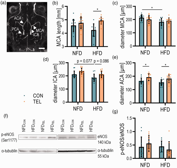
Figure 4.
TEL affects cerebral vessel diameter upon HFD. Mice were fed with normal-fat (NFD) or high-fat diet (HFD) for 16 weeks and simultaneously treated with vehicle (CON) or telmisartan (TEL). (a) Representative image of TOF-angiography and analyzed vessels (indicated by arrows; ACA anterior cerebral artery; ICA internal carotid artery; MCA middle cerebral artery; PCA posterior cerebral artery; A anterior; P posterior; L left; R right; scale bar 2 mm) with (b) length of MCA, diameter of (c) MCA, (d) ICA and (e) ACA are shown. (f) Representative western blot images and (g) quantification of p-eNOS/eNOS ratio. 2-way ANOVA results see table S5. *P < 0.05 in Bonferroni multiple comparison posttest; (a)–(e) n = 12–13 per group, (f)–(g) n = 10 per group; Data are meansSD.

