Figure 6.
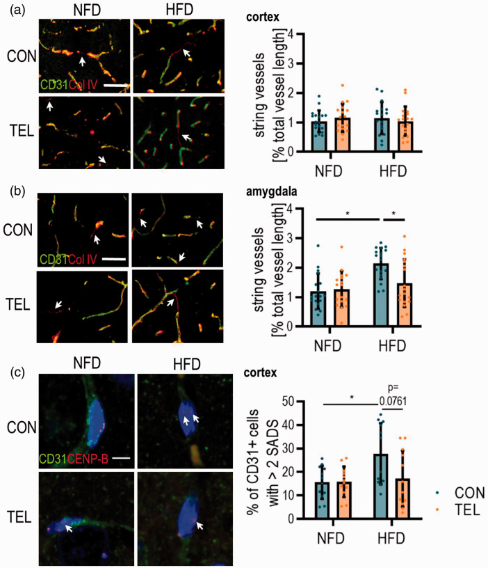
TEL has protective effects on cerebral vasculature morphology. Mice were fed with normal-fat (NFD) or high-fat diet (HFD) for 16 weeks and simultaneously treated with vehicle (CON) or telmisartan (TEL). Representative images of immunohistochemistry with antibodies for a marker of endothelial cells (CD31) and basal membrane (Col IV) and quantification of string vessel length (CD31−/Col IV+, indicated by arrows) in (a) cortex and (b) amygdala; scale bar 50 µm. (c) Representative images showing peri/centromeric satellite DNA signals in the cortical area (SADS, indicated by arrows); Micrographs showing single CD31+ (green) cell nuclei with centromere protein-B (CENP-B, red); scale bar 5 µm and percentage of CD31+ cells with >2 SADS are shown (right). 2-way ANOVA results see table S7. *P < 0.05 Bonferroni multiple comparison posttest. (a)–(b) n = 17–18 per group (4 images per n) and (c) n = 6 per group (in average 52 CD31+ cells per n); Data are meansSD.

