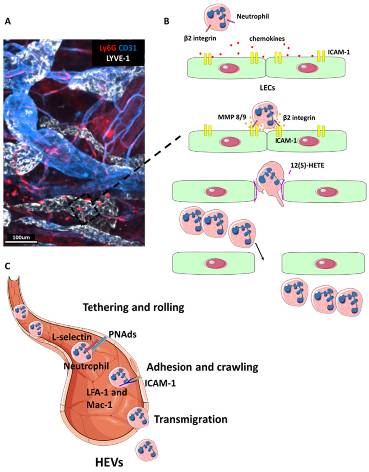
Figure 3.
Neutrophil migration to lymphoid organs. (A) Two-photon microscopy image of a section of inflamed skin in a Ly6G-Tomato reporter mouse [48] stained with LYVE-1 (white) to visualise LECs and endothelial cells with CD31 (blue). Neutrophils (red) can be seen in and around lymphatic vessels. (B) Schematic representation of neutrophil transmigration across lymphatic endothelium. First, neutrophils chemotax toward LECs following inflammation-induced release of chemokines such as CXCL8 in humans and CXCL1 in mice [49]. Next, neutrophils adhere to inflamed LECs via integrin binding to ICAM-1. This triggers the secretion of MMP8, MMP9 and exocytosis of chemorepellent 12(S)-HETE, which together promote transient junctional retraction and neutrophil transmigration. The resulting gap in the lymphatic endothelium serves as a hotspot for rapid neutrophil transmigration. Adapted from D.G. Jackson, Frontier Immunology, 2019 [2]. (C) Diagram showing neutrophil entry into the lymph node via HEVs. Neutrophil express L-selectin which binds to glycoproteins expressed on the HEVs walls, such as peripheral node addressins (PNAds) to allow tethering and rolling, which promote rapid adhesion and emigration [15]. Neutrophil adhesion is mediated via binding of ICAM-1 to integrins LFA-1 and Mac-1. In particular, LFA-1 mediates neutrophil adhesion and Mac-1 facilitates subsequent crawling [50].

