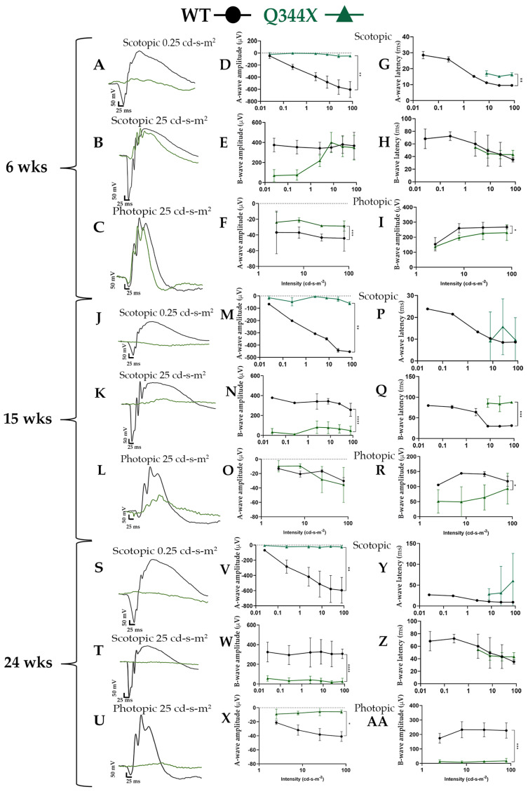
Figure 1.
Q344X rhodopsin knock-in mice elicit minimal rod cell responses by ERG. (A–C, J–L, S–U) Overlaid representative scotopic and photopic ERG traces at low and high flash intensities (0.25 and 25 cd-s-m2) from dark and light-adapted animals at 6 weeks (A–C), 15 weeks (J–L), and 24 weeks (S–U): WT (black) and Q344X (green). (D–AA) Summary data averaged across WT (black circles) and Q344X (green triangles). A-wave and b-wave amplitudes were measured from base line to trough or peak respectively for each flash intensity for 6 weeks (D–F), 15 weeks (M–O), and 24 weeks (V–X). Latencies represent time to peak or trough for each flash intensity for 6 weeks (G–I), 15 weeks (P–R), and 24 weeks (Y–AA). Q344X mice demonstrated significantly decreased a-wave amplitude compared to WT between 3 and 6 weeks. A measurable b-wave was present in scotopic tracings of young Q344X mice; however, the tracing correlated with b-waves in photopic tracing. At 24 weeks, Q344X mice responses were flatline (n = 3-4 per group; mean ± SD; * p = 0.02, ** p < 0.05, *** p < 0.0005, **** p < 0.0001).

