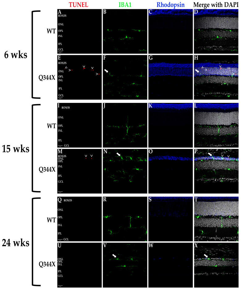
Figure 4.
Photoreceptors undergo apoptosis while microglia proliferate and phagocytose non-apoptotic cells in the Q344X rhodopsin knock-in mouse retina. WT (A–D, I–L, Q–T) and Q344X (E–H, M–P, U–X) mouse retinal sections at 6, 15, and 24 weeks of age were TUNEL-labeled (red) and immunolabeled for IBA1 (green) and rhodopsin (blue). Nuclei were labeled with DAPI (white). ROS/IS, rod outer segments/inner segments; ONL, outer nuclear layer; OPL, outer plexiform layer; INL, inner nuclear layer; IPL, inner plexiform layer; GCL, ganglion cell layer. Arrowheads (>) indicate apoptosing cells. Arrows indicates phagoptosis (aberrant phagocytosis of living cells by microglia). Scale bars = 20 µm.

