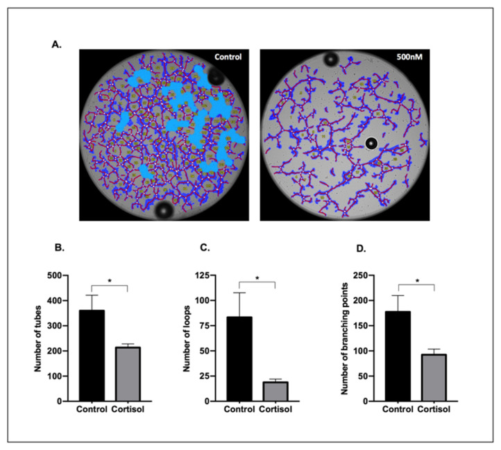
Figure A1.
Tube-like structures formation—a pilot experiment. Representative images of tube formation (A) and the quantification of the number of tubes (B), loops (C), and branching points (D) of SGHPL-4 cells treated with vehicle (0 nM) or cortisol (500 nM) in the medium. Images were obtained after 5 h of stimulation. Data were compared with t-test and Shapiro–Wilk test; * p < 0.05. Data are presented as mean ± SEM. The quantification of tubes, loops, and branching points was done with the WimTube Software (Wimasis, Munich, Germany).

