Figure 3.
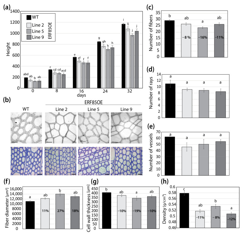
Ectopic expression of PtERF85 in woody tissue increased fibers diameter and reduced cell wall thickness. (a) Stem growth rate of WT and three transgenic lines that ectopically express PtERF85 (ERF85OE) under a xylem specific wood promoter (pLMX5; [15]) in a controlled greenhouse environment. (b) Transmission electron micrographs (TEM; 100-fold magnification; scale bar is 10 µm) and toluidine blue staining (20-fold magnification; scale bar is 40 µm). TEM pictures cover a total area of 149,295 µm2. (c) The number of fiber cells counted in toluidine blue cross-sections is shown in (b). (d,e) Numbers of ray and vessel cells (in an area of 3.02 mm2) in WT and ERF85OE lines. Cross-sections used for quantification are shown in Figure S3b. (f) Fiber diameter from toluidine blue-stained cross-sections is shown in (b). Cell outlines were defined by the middle lamella and the enclosed area [µm2] was measured using ImageJ. (g) Cell wall thickness (determined as the lumen-to-lumen distance of two neighboring cells [µm]) based on toluidine blue-stained stem sections (shown in (b)), using a 100-fold magnification. (h) Wood density. For all panels, bars represent mean ± SE calculated from three biological replicates per line using a linear effect model. Significant differences between genotypes were indicated by the unique occurrence of letters above the bars and were assigned based on multiple comparison tests and a p-value < 0.05.

