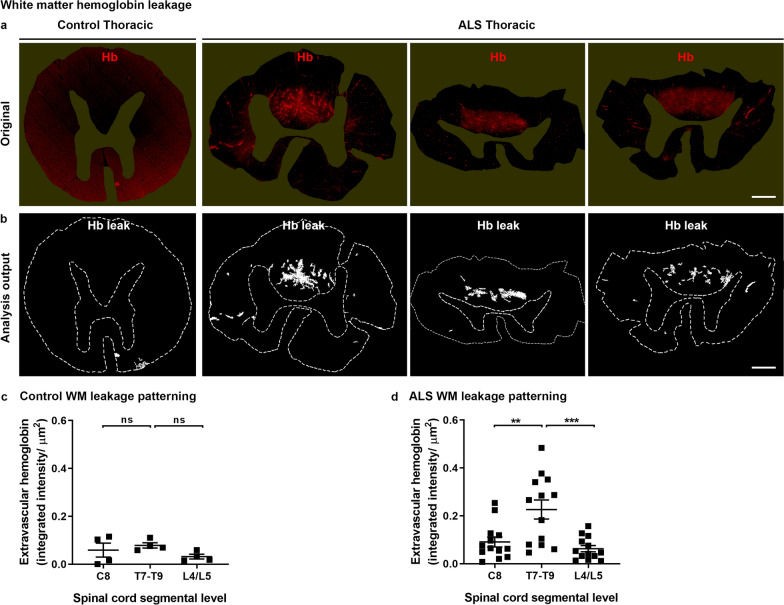
Fig. 5.
White matter hemoglobin leakage along the spinal cord axis. Immunohistochemical labelling (a) and automated analysis outputs (b) of extravascular hemoglobin in control and ALS spinal cord white matter. Scale bars = 1 mm. Quantification of extravascular hemoglobin in gray matter at individual segmental levels C8, T7–T9 and L4/L5 in control (c) and ALS (d) cases. Data shown as mean ± SD (control n = 4, ALS n = 13), with statistical significance determined using two-way ANOVA with Tukey’s post-test. ***p ≤ 0.001; **p ≤ 0.01; ns, not significant

