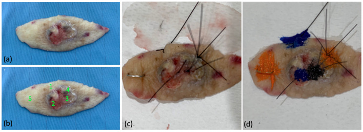
Figure 2.
Example of lesional and healthy skin reference points on excised specimen: (a) specimen after excisional biopsy; (b) ex vivo identification of control points (1,5) and lesional points (2,3,4) for 400–430 nm irradiation; (c) suture stitches as reference for histologic analysis, (d) tissue marking dyes before histopathological examination.

