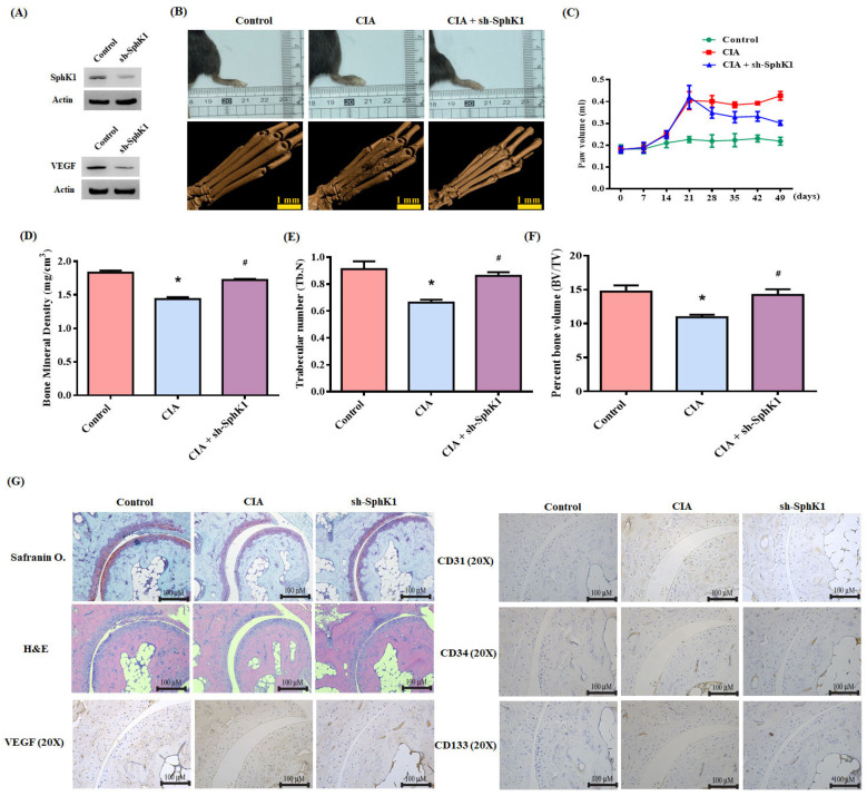
Figure 7.
S1P knockdown reduces the in vivo severity of RA. (A) After infecting osteoblasts with control or SphK1 shRNA, Western blot determined SphK1 and VEGF expression. CIA mice received intra-articular injections of ~7.1 × 10⁶ PFU SphK1 shRNA or control shRNA on day 14 and were sacrificed on day 49. (B) Representative µ-CT images of the hind paws taken on day 49. (C) A digital plethysmometer quantified the amounts of hind paw swelling. (D–F) µ-CT SkyScan Software quantified BMD, trabecular numbers and bone volume. (G) Histological sections of ankle joints were stained with H and E or Safranin O/fast green, and immunostained with VEGF, CD31, CD34 and CD133. Results are expressed as the mean ± S.D. (n = 3). * p < 0.05 versus the control group; # p < 0.05 versus S1P alone.

