Figure 1.

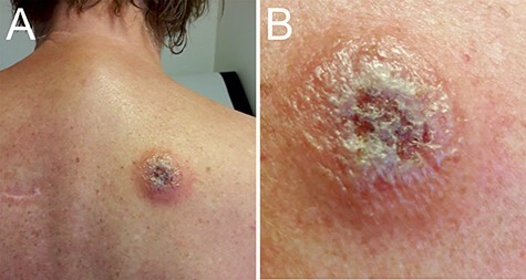
Trypanosomal chancre. Panel A shows the trypanosomal chancre on the back of the patient at presentation. The chancre had a diameter of ca. 5 cm with ulcerating and elevated edges. Panel B shows the swollen chancre at higher magnification.

Trypanosomal chancre. Panel A shows the trypanosomal chancre on the back of the patient at presentation. The chancre had a diameter of ca. 5 cm with ulcerating and elevated edges. Panel B shows the swollen chancre at higher magnification.