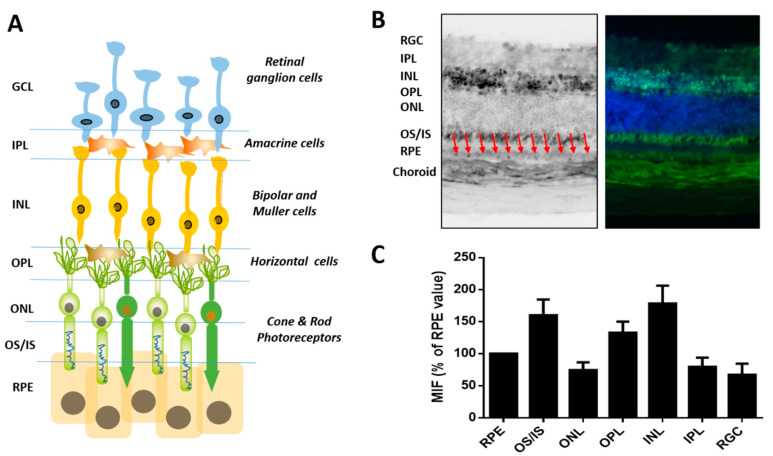
Figure 4.
Immunohistochemistry of GLO1 in mouse retinal tissues. (A) Cross-sectional, cellular schematic of the retina illustrating its three primary layers comprised of the ganglion cell layer (GCL), containing retinal ganglion cells (RGC), inner nuclear layer (INL), hosting interneurons of amacrine, bipolar and horizontal cells as well as Müller glial cells, and outer nuclear layer (ONL), housing rod and cone photoreceptors. The sensory tissue, or neuroretina, is connected to the retinal-pigmented epithelium (RPE). Red arrows indicated the RPE layer. (B) Representative picture of GLO1 immunostaining in retinal samples from WT mice. (C) Mean intensity fluorescence of GLO1 normalized to the value in the RPE. Data shown are mean ± standard errors of the means (SEM).

