Figure 1.
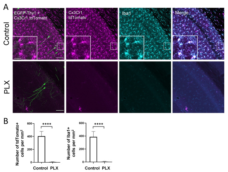
PLX treatment for 28 days leads to microglia depletion. (A) The confocal images show perfect colocalization of tamoxifen-induced tdTomato (magenta) signal in Cx3Cr1-expressing cells and immunohistochemical staining for microglial marker Iba1 (cyan) in the CA1 region of the hippocampus. Insets illustrate the typical microglial morphology of labeled cells. EGFP labels a small subset of pyramidal neurons (green), which were used for dendritic spine analysis. (B) The bar graphs display the efficacy of the oral PLX3397 administration. After 4 weeks of treatment, virtually all microglia are depleted from the hippocampus. **** p <0.0001, t-test. Scale bars are 100 µm for main images and 25 µm for inserts.

