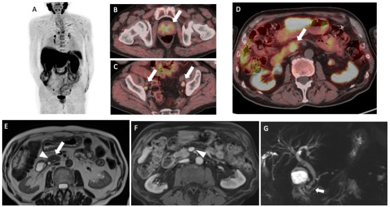
Figure 1.
A male patient with prostate cancer underwent 18F-Choline (FCH) PET/CT for the suspicion of lymph node recurrence during hormonal therapy. He was not submitted to local therapies (i.e., surgery or radiotherapy) for the treatment of the primary prostate lesion. The patient has, in addition, a history of diabetes mellitus type II and hypertension. PET/CT detected multiple areas of focal FCH uptakes ((A); maximum intensity projection—MIP) in prostate gland (arrow, (B)) and abdominal-pelvic lymphadenopathies (arrows, (C)), compatible with recurrent prostate cancer. Moreover, a focal tracer uptake was shown in the uncinate process of the pancreas with a maximum standardized uptake value (SUVmax) equal to 13.6 (arrow, (D)). As already stated by Schillaci et al. [1], FCH may physiologically show a moderate-to-high uptake in the liver and the pancreas. However, the presence of a focal uptake should be further investigated in order to make a differential diagnosis with a malignant or benign lesion. A subsequent MRI showed no definite lesion but rather a slight “mass-like” enlargement of the pancreatic head, as visible on the axial single-shot turbo-spin echo T2-weighted image (arrow, (E)). Some micro-cystic areas were appreciated in the pancreaticoduodenal groove (arrowhead, (E)). The absence of lesions was confirmed on the post-contrast pancreatic phase (axial gradient echo volumetric fat saturated image), in which pancreatic parenchyma appears homogeneous, with no solid tissue extending towards major vascular structures (arrowhead, (F)). Of note, the cholangiopancreatography sequence showed a normal-sized but irregular main pancreatic duct, with no cephalic structures (arrow, (G)). Overall, subtle morphological changes and ductal abnormalities supported the diagnosis of CMFP. The Carbohydrate antigen 19–9 (CA19.9) was normal (17.0 UI/mL). However, after one month from MRI and FCH PET/CT, the patient was submitted to an endoscopic ultrasound (EUS) exam and biopsy, based on the suggestion of the multidisciplinary team.

