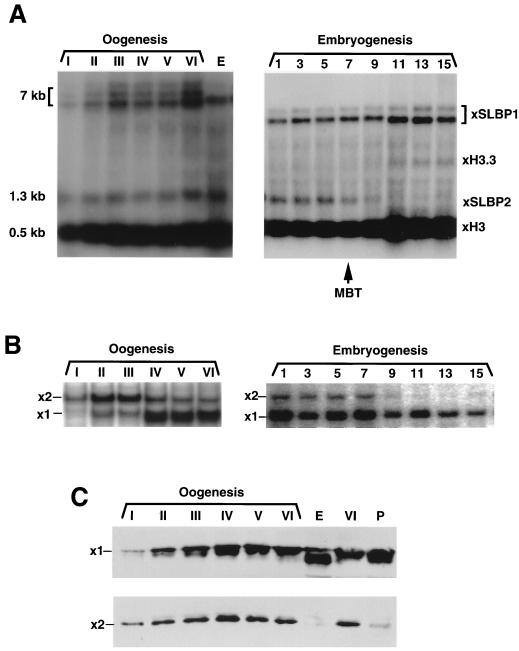
FIG. 5.
Expression of SLBP1 and SLBP2 during oogenesis and early embryogenesis. (A) RNA was prepared from oocytes and early embryos and resolved by gel electrophoresis on a 1% agarose gel. RNA from two oocytes or embryos was analyzed. The gel was transferred to nitrocellulose and hybridized with a mixture of probes to SLBP1, SLBP2, and histone mRNA. The histone probe was made at one-half of the specific activity of the other two probes. The identity of the bands was confirmed by hybridizing with single probes. (B) Extracts were prepared from oocytes and embryos and assayed for SLBP1 and SLBP2 by mobility shift assay. Each lane is analysis of one oocyte. (C) Protein from one oocyte (top) or four oocytes (bottom) was analyzed by Western blotting with either affinity-purified anti-SLBP1 (top) or affinity-purified anti-SLBP2 (bottom). Lanes P show oocytes matured by treatment with progesterone, and lanes E show eggs. The apparent higher mobility of SLBP1 in eggs in this experiment was not seen in other experiments.

