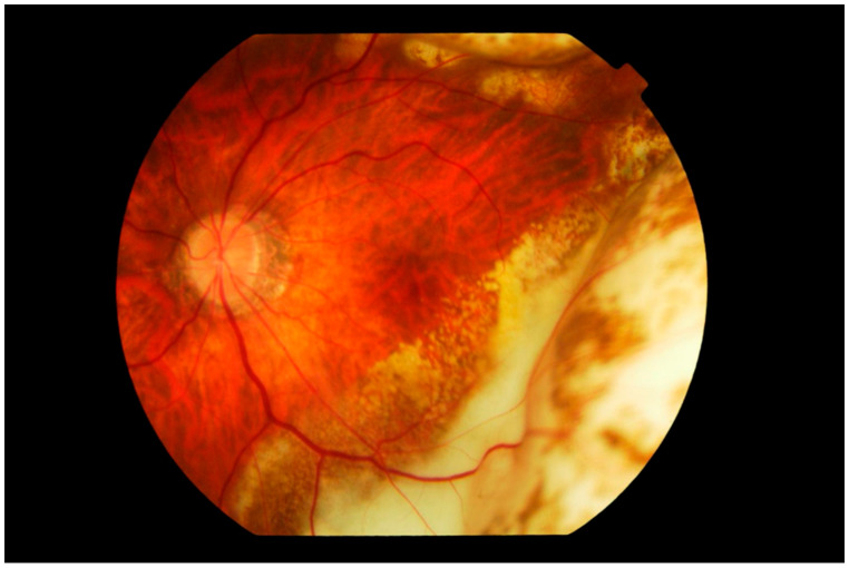
Figure 3.
Fundus photograph of the left eye showing subretinal infiltration by lymphoma cells forming multiple large yellowish cream nodular masses distributed circumferentially with patchy pigmentation, giving a characteristic “leopard spot” appearance. The inferior temporal fundus shows the recent development of a wide span of subretinal infiltrates, contiguous with the longer standing peripheral mass. The edge of the lesion advancing towards the fovea consists of multiple small cream-colored spots. The vitreous is clear.

