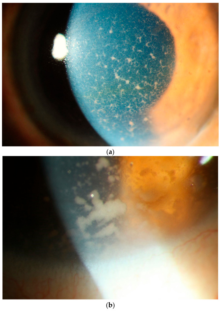
Figure 7.
Slit lamp photograph showing (a) diffusely distributed keratic precipitates (KP) on the corneal endothelium. These KPs are a mixture of small and fine or infiltrative KP intermixed with some granulomatous KP. Many of these granulomatous KP have fibrillar extensions, taking on a comet-like appearance, typical of vitreoretinal lymphoma. These faintly pigmented KP may be mistaken for the KP of viral anterior uveitis. (b) Occasionally one can find even larger tumor cell collections on the endothelium.

