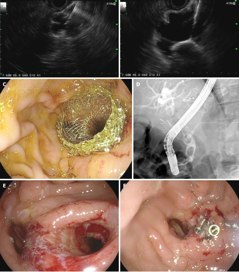
Figure 4.
Endoscopic ultrasound-directed transgastric endoscopic retrograde cholangiography for choledocholithiasis in a patient with history of roux-en-Y gastric bypass surgery. A: Endoscopic ultrasound-guided puncture of excluded stomach using a 19-gauge needle; B: Endoscopic ultrasound showing deployment of proximal flange of lumen-apposing self-expanding metal stent (LAMS) in the excluded stomach; C: Endoscopic image showing distal flange of LAMS in the gastric pouch; D: Fluoroscopic image of endoscopic retrograde cholangiopancreatography through LAMS showing multiple stones in the common bile duct; E: Gastrogastric fistula seen following LAMS removal; F: Successful closure of gastrogastric fistula using argon plasma coagulation and clips.

