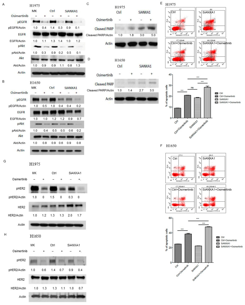
Figure 4.
Western blot analysis of EGFR, pEGFR (Tyr1068), Akt (Ser473), pAkt and actin in (A) H1975 and (B) H1650 lung cancer cells transfected with 50 nM scrambled control (Ctrl) and ANXA1 (SiANXA1) siRNA for 72 h and then treated with 10 nM Osimertinib for 3 h. MK: mock, without transfection of siRNA. The relative expression of pEGFR, EGFR, pAkt and cleaved PARP were normalized to actin using MK group as the control. MK: mock; pEGFR: phosphor EGFR; pAkt: phosphoAkt. Western blot analysis of cleaved PARP in (C) H1975 and (D) H1650 lung cancer cells transfected with 50 nM scrambled control (Ctrl) and ANXA1 (SiANXA1) siRNA for 72 h and then treated with or without 100 nM Osimertinib for 24 h. Actin was used for internal control. The relative expression of ANXA1 was normalized to actin using Ctrl group as the control. Apoptosis assay by Annexin V-FITC in (E) H1975 and (F) H1650 lung cancer cells after transfection of 50 nM scrambled control (Ctrl) or ANXA1 (SiANXA1) siRNA with or without 100 nM Osimertinib for 24 h. The percentage of apoptotic cells is represented as bar ± standard deviation in triplicate experiments. Western blot analysis of HER2, pHER2 (Tyr877) and actin in (G) H1975 and (H) H1650 lung cancer cells transfected with 50 nM scrambled control (Ctrl) and ANXA1 (SiANXA1) siRNA for 72 h and then treated with 10 nM Osimertinib for 3 h. The relative expression of pHER2 and HER2 were normalized to actin using MK group as the control. MK: mock; pHER2: phosphor HER2. “ns” denotes nonspecific. “***” denotes p < 0.001. Uncropped western blot figures were included in Supplementary Materials.

