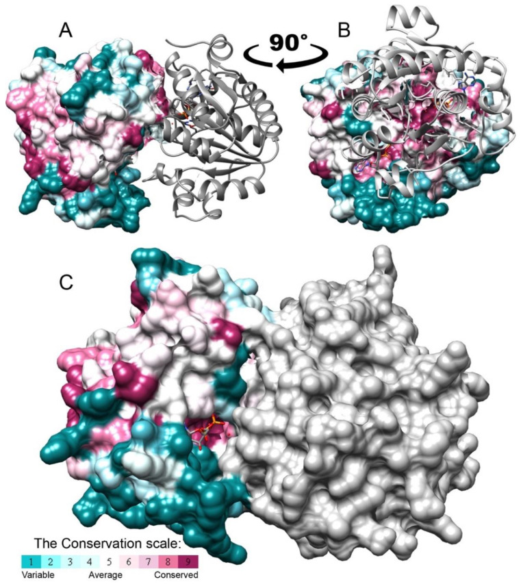
Figure 6.
The isolated BceF kinase domain forms a dimer in the crystal structure. A dimer of the BceF kinase domain was observed in the crystal structure, demonstrating a partial obstruction of the active site. One monomer is colored grey and the other by evolutionary conservation scores calculated using the ConSurf web server [21,22,23]; a variable-to-conserved colored scale is indicated. ADP, present in the crystal structure, is shown in format of sticks and colored by atom type. (A,B) Two views (representing −90° rotation along the vertical direction) of the BceF dimer with one monomer displayed as ribbons and the other as solvent-accessible surface representation. (C) The two monomers are showed using a solvent-accessible surface representation.

