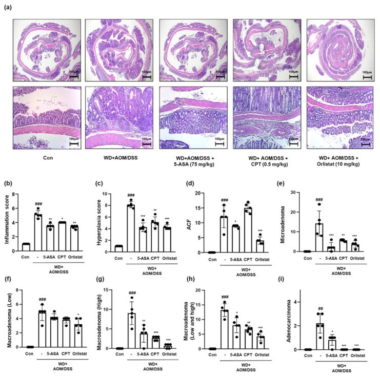
Figure 4.
Effect of orlistat on the histological pathology of CAC in WD-driven CAC mice. (a) H&E staining was conducted using colon tissues from each group. Slide sections were scrutinized by microscopy. (b) Inflammation scores; (c) hyperplasia scores; (d) ACF scores; (e) microadenoma scores; (f) macroadenoma (low); (g) macroadenoma (high); (h) macroadenoma (low and high); (i) adenocarcinoma values. The extent of histological pathology was evaluated by a medical technologist. The values are the means ± SD (n = 5); ## p < 0.01 and ### p < 0.001 when compared to Con; * p < 0.5, ** p < 0.01, and *** p < 0.001 when compared to WD + AOM/DSS.

