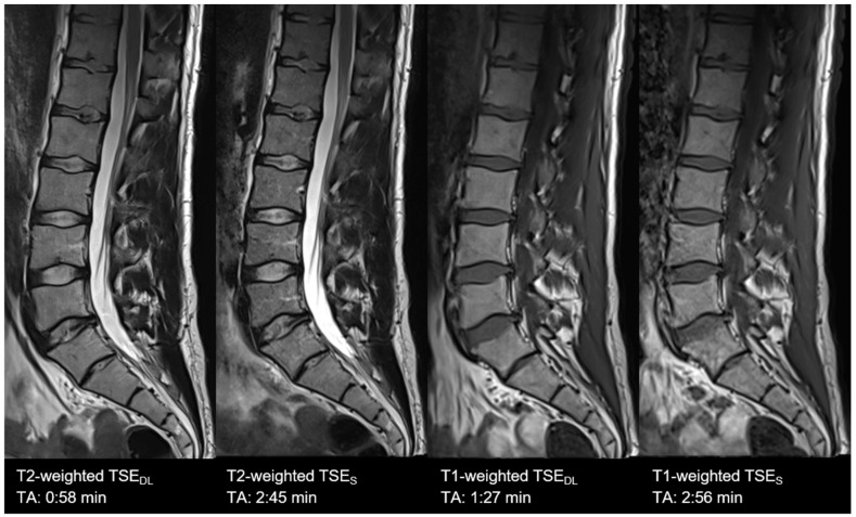
Figure 5.
Example of a Deep Learning and standard T1- (right) and T2-weighted (left) turbo spin echo image of the lumbar spine in sagittal orientation. Note that TSEDL show lower extents of noise both in T1- and T2-weighted imaging. Nonetheless, some small structures, such as small bone canals, disappear; there is no impact on the delineation and assessment of relevant anatomical structures in both TSES and TSEDL.

