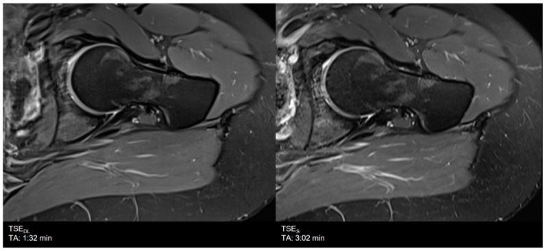
Figure 6.
Example of a Deep Learning (left) and standard (right) PD-weighted turbo spin echo image of the hip in axial orientation. Note that although the assessment of the bone was rated to be lower, the assessment of anatomical structures and articular cartilage, as well as the delineation of ligaments and tendons, are comparable between TSEDL and TSES.

