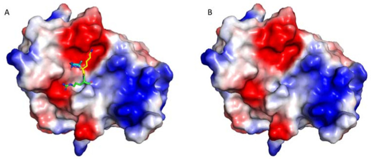
Figure 3.
The protease active site is negatively charged. The structures of eZiPro (PDB ID 5GJ4) with (A) and without (B) TGKR sequence of NS2B are shown to understand the surface charges. The TGKR residues are shown in different color. The surface charge figure was made using PyMOL (www.pymol.org (accessed on 3 August 2018)). Surface areas with positive charge, negative charge, and no charge are shown in blue, red, and white, respectively. The substrate-binding site is negative charged, suggesting the challenges of developing small molecule inhibitors, which prefer interacting with a hydrophobic surface.

