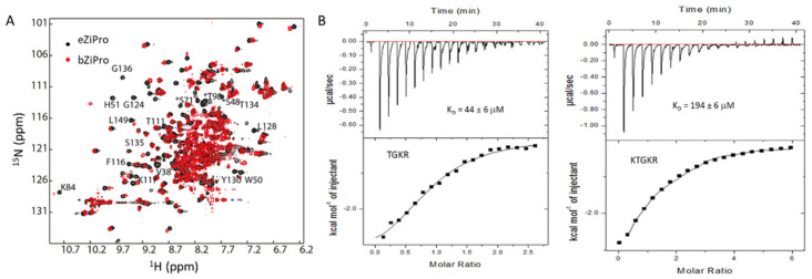
Figure 4.
Dynamics and conformational changes in ZIKV protease. (A). Overlay of 1H-15N-HSQC spectra of eZiPro and bZiPro. This figure is obtained from the reference [58]. The 1H-15N-HSQC spectra of eZiPro and bZiPro are shown in black and red, respectively. More cross-peaks appeared in eZiPro suggests that substrate binding to protease suppresses exchanges. (B). Binding affinity between protease and peptides. The weak binding affinity is important for the function of the protease. This figure was obtained from the reference [108] with permission.

