Figure 1.
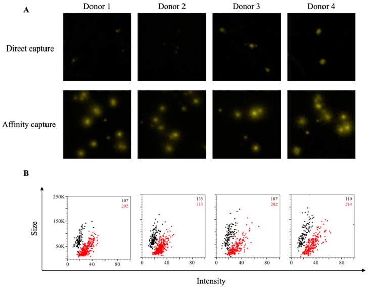
Affinity tag capture improves detection of SARS-CoV-2 RBD-reactive ASC. PBMC from four PCR-confirmed COVID-19 donors were stimulated in vitro (detailed in Materials and Methods) and evaluated for antibody-secreting (ASC) reactivity against the receptor-binding domain (RBD) fragment of the SARS-CoV-2 Spike protein. (A) Representative well images depicting antigen-specific IgG+ ASC in wells coated directly with 10 μg/mL of RBD protein or through affinity capture using the genetically encoded hexahistidine (6XHis) tag. Magnification and contrast enhancements were uniformly performed on all images to aid their visualization in publication. (B) RBD-specific FluoroSpots were merged into flow cytometry standard (FCS) files (detailed in Materials and Methods) and visualized as bivariate plots measuring spot intensity (x-axis) and spot size (y-axis). FluoroSpots originating from assay wells in which RBD protein was directly captured on the membrane (black dots) or through affinity capture (red) are shown as overlays. The combined number of FluoroSpots (spot-forming units, SFU) detected in replicate wells for each of the respective donors is indicated in the inset using the same red/black color code.

