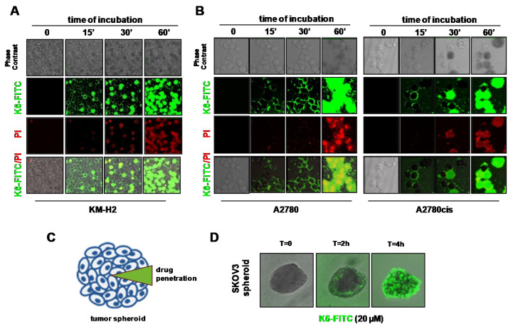
Figure 4.
K6-FITC uptake by HL OvCa cells and OvCa spheroids. Representative confocal images showing (A) KM-H2 (B) A2780 (left panels) and A2780cis cells (right panels) double stained with K6-FITC (30 µM) (green) and propidium iodide (PI) (red) at various time points. (C) Illustration of drug penetration kinetic into spheroid. (D) Representative photomicrographs of a SKOV3 spheroid treated with K6-FITC (20 µM) and analyzed for K6-FITC uptake by confocal microscopy time-lapse.

