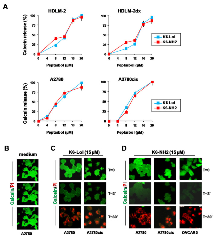
Figure 6.
K6-Lol and K6-NH2 rapidly induce calcein release and cell death in OvCa cells. Membrane permeability assays. (A) To monitor the lack of the cell membrane integrity after peptaibols treatment, tumor cells were prestained with calcein AM. Then, cells were treated with peptaibols (0–20 µM) and fluorescent calcein in supernatants was evaluated immediately (2 min). Results are expressed as the percentage of calcein released by cancer cells with respect to cells disrupted with triton X (100%, total fluorescent calcein converted to a green fluorescent form by cancer cells). Bar charts are mean and SD of three independent experiments. (B–D) Cell-based calcein release assay evaluated by confocal microscopy. OvCa cells were preincubated with calcein AM (green staining), then stained with PI (to evaluate dead cells) (red staining) and treated with peptaibols. (B) Then, untreated cells (medium) and (C,D) cells treated with peptaibols (15 µM) were monitored for calcein AM release by real-time fluorescence using the Operetta system.

