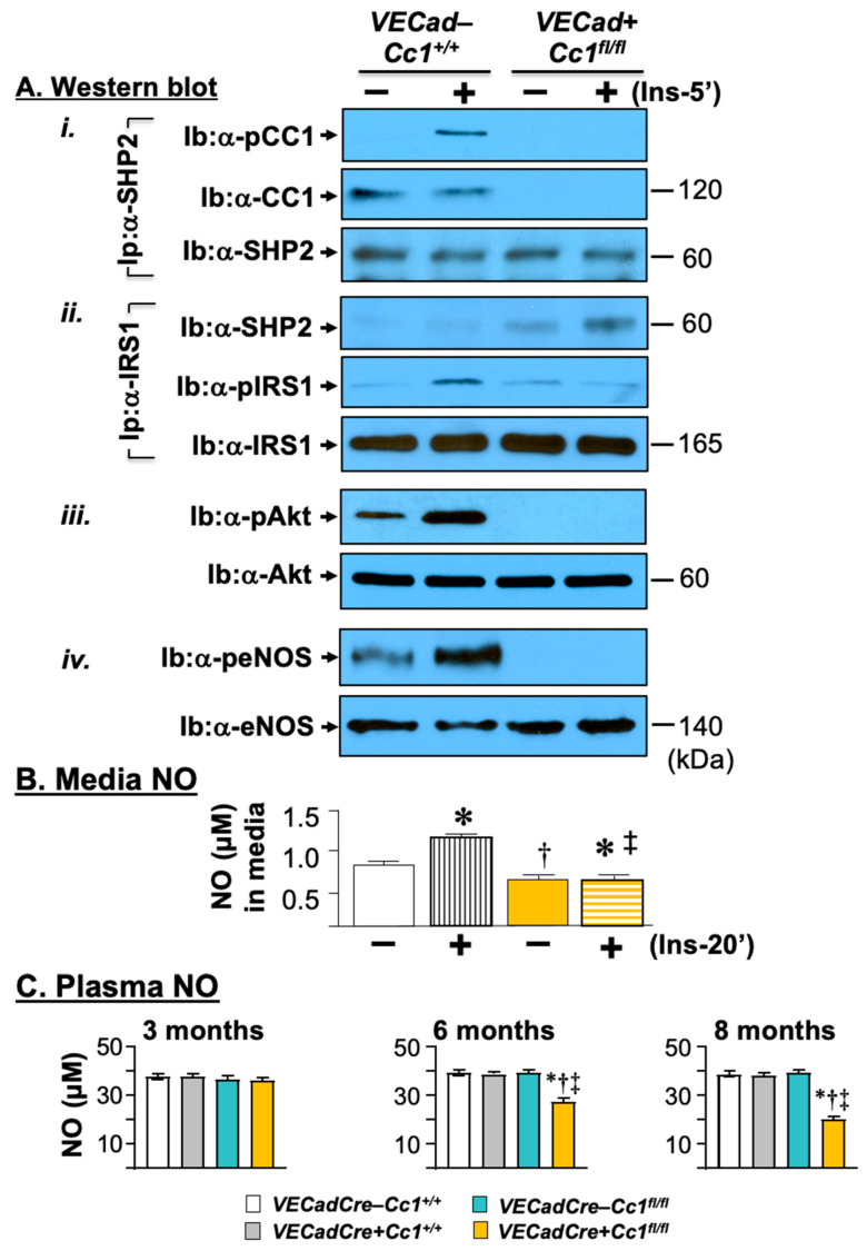
Figure 5.
Insulin signaling leading to nitric oxide production in liver endothelial cells. (A) Primary liver endothelial cells (LEC) from 2-month-old WT (VECad−Cc1+/+) and null (VECad+Cc1fl/fl) mice were treated with (Ins, 100 nM) or without insulin for 5 min before proteins were lyzed and (i) subjected to immunoprecipitation (Ip) with SHP2 antibody followed by immunoblotting (Ib) with antibodies against phospho-CEACAM1 (pCC1) and CEACAM1 (CC1) or SHP2 to assess the amount of CC1 and pCC1 in the SHP2 immunopellet. (ii) Similar co-immunoprecipitation experiments were performed to evaluate the association between IRS1 with SHP2. (iii,iv) Lysates were also subjected to immunoblotting with α-phospho-antibodies to account for activation of Akt and eNOS, respectively, as in the legend in Figure 4. Gels represent two separate experiments. (B) levels of nitric oxide (NO) were determined in triplicate in the media of cells treated with or without insulin for 20 min. Values are expressed as mean ± SEM. * p < 0.05 vs. no insulin/same genotype; † p < 0.05 vs. VECadCre−Cc1+/+ no insulin; ‡ p < 0.05 vs. VECadCre−Cc1+/+ plus insulin.(C) Male mice (3–8 months of age, n ≥ 5/genotype) were fasted overnight before blood was drawn, and plasma was processed to assess NO levels. Values are expressed as mean ± SEM. * p < 0.05 vs. VECadCre−Cc1+/+, † p < 0.05 vs. VECadCre+Cc1+/+, ‡ p < 0.05 vs. VECadCre−Cc1fl/fl.

