Figure 3.
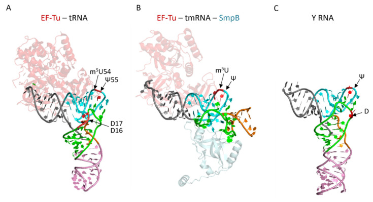
The structures of tmRNA and Y RNA mimic part of the tRNA structure. (A) The structure of EF-Tu-tRNAPhe complex (with GDPNP, GTP analog) from Thermus aquaticus (pdb file 1TTT [189]). Each domain of the tRNA is colored: acceptor am in grey, TY-arm in light blue, the variable region in orange, the D-arm in green, and the anticodon-arm in pink. EF-Tu structure is represented in light pink. (B) The structure of the tmRNA fragment in complex with EF-Tu (with GDP and kirromycin antibiotic, in light pink) and SmpB (in light grey) from Thermus thermophilus (pdb file 4V8Q [190]). The regions of tmRNA mimicking tRNA are shown with the same color code as for the tRNA. (C) Salmonella Typhimurium Y RNA (YrlA, pdb file 6CU1 [191]). The regions of YrlA mimicking tRNA are shown with the same color code as for the tRNA.

