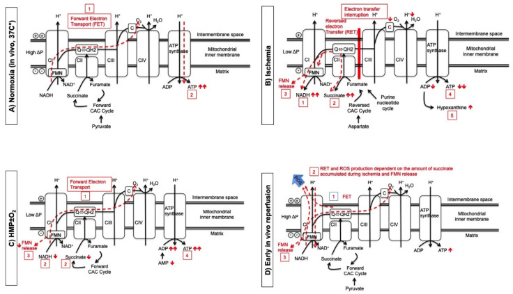
Figure 3.
The effect of active oxygenation during HMP on mitochondria through the process of kidney preservation and transplantation. (A) During normoxia, forward electron transport (dotted line) (1) drives the proton pumps (CI, CIII, CIV) against a charge gradient at the oxidative phosphorylation chain and proton back flow drives ATP synthase to generate ATP (dotted line) (2). (B) In the absence of oxygen, electron transfer is interrupted and first NADH (1) and second citric acid metabolite succinate (2) increase because both act now as electron carrier to allow CI continuously pump protons during ischemia. Also FMN release (3) at CI results from over-reduction of flavin via RET. Consequently, ATP and ADP decrease (4) and purine metabolites increase (5). (C) The degree of FET during HMP depends on the perfusate oxygen concentration (1) and results in a decrease of succinate and NADH (2) and less FMN release (3) and an increase of ADP/ATP regeneration (4). (D) During early, in vivo reperfusion, FET (1) generates ATP. Rapid consumption of accumulated succinate generates too much QH2, which impairs further FET. In combination with acidic pH from ischemia this results in a RET at CI (2). Reduced or semi-reduced FMN (3) located at the nucleotide-binding site is the direct source of ROS in CI during RET but reduced FMN can also non-enzymatically react with molecular oxygen producing ROS.

