Figure 1.
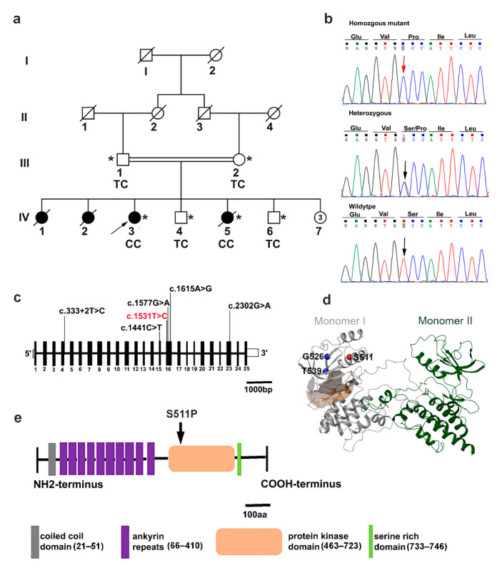
Pedigree, sequencing chromatogram, and location of the TNNI3K variant in the family with cardiac conduction disease (CCD). (a) Family pedigree. The open symbols represent unaffected individuals and the filled symbols represent affected individuals; symbols with a diagonal line represent deceased individuals; and the arrowhead designates the proband. An asterisk indicates family members from whom DNA was available. The genotypes of the TNNI3K mutation are indicated below each examined member: CC (homozygote); TC (heterozygote); number inside the circle denote the number of individuals. (b) Sequencing chromatograms. Vertical arrows indicate the mutation site. (c) Schematic of the human TNNI3K gene. The positions of coding exons (black) and UTRs (white) are indicated. Black arrows indicate previously reported pathogenic variants and the red arrow shows the novel variant c.1531T>C in exon 16. (d) Initial 3D structure of TNNI3K-WT (PDB-ID: 4YFI [29]) shown as a ribbon. Monomer I is shown in gray and monomer II in green. The Cα atoms of Ser511 (red), Gly526 (blue), and Thr539 (blue) of monomer I are shown in spheres relative to the ATP-binding pocket (orange surface) of monomer I. Gly526 and Thr539 are known TNNI3K missense variants. (e) TNNI3K protein domain structure. Coiled coil domain (gray); functional ankyrin repeat domains, ANK1–ANK10 (purple); kinase domain (orange), where the homozygous mutation p.Ser511Pro resides (arrowed); and a serine-rich domain (spring green).

