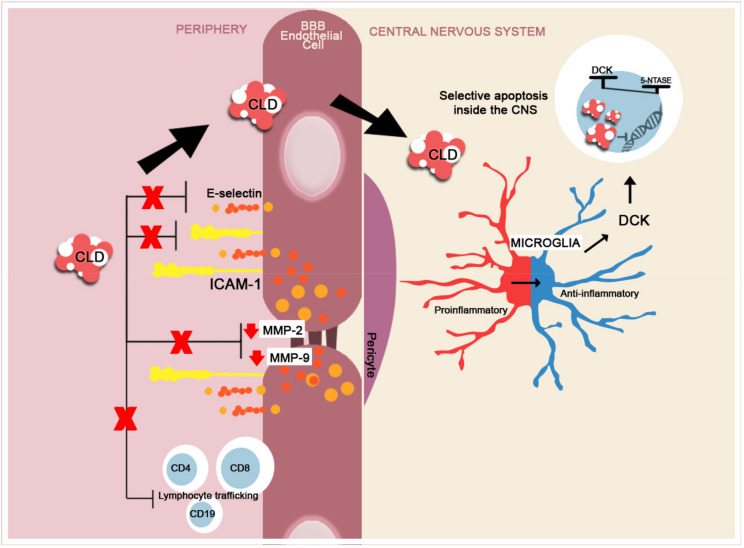
Figure 5.
A brief schematic representation of CLD’s mechanism of action. In the peripheral compartment, CLD reduces the expression of adhesion molecules, ICAM-1 and E-selectin and reduces the expression of MMP-2 and -9 [136,137,138,139], thus inhibiting the lymphocyte transition into the CNS. CLD is internalized into the cells and undergoes specific phosphorylation by deoxycytidine kinase (DCK). Immune cells, the lymphocytes being the most susceptible, contain reduced levels of phosphatases 5-nucleotidases (5-NTASE), which will only partially dephosphorylate the accumulated CLD, leading to selected apoptosis and not cellular death (ratio of DCK to 5-NTASE) [140,141]. In MS, CLD selectively reduces Th and B lymphocyte numbers and trafficking from the periphery [142]. CLD reduces the activation of proinflammatory microglia (M1) and shifts the activity towards an anti-inflammatory (M2) phenotype [138].

