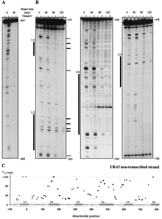
FIG. 2.
Repair of UV-induced CPDs at single nucleotide resolution along (A) the transcribed strand, nt 268 to 607 (all positions are relative to the start codon, ATG, designated +1), and (B) the nontranscribed strand, nt −151 to 221, nt 324 to 518, and nt 478 to 760. Cells were irradiated with 70 J/m2, and repair was monitored at 0, 40, 80, and 120 min after irradiation. Samples that were mock treated or treated with the CPD-specific enzyme T4endoV are denoted by − and +, respectively. Shaded boxes indicate the internal protected regions of nucleosomes U1, U2, U4, and U5 positioned along the URA3 locus (19). Dark arrows mark CPDs that persisted after 2 h of repair, and open arrows mark some positions that were repaired very fast. (C) Graphic representation of quantified CPD repair rates along the nontranscribed strand of the URA3 locus. Repair t1/2 values, determined as the time at which 50% of the initial CPD signal was removed, were calculated for each individual CPD position with a sufficient signal-to-noise ratio and are plotted above their corresponding dipyrimidine positions. The internal protected regions are represented by the shaded boxes inside nucleosomes U1 through U6 (19).

