Figure 7.
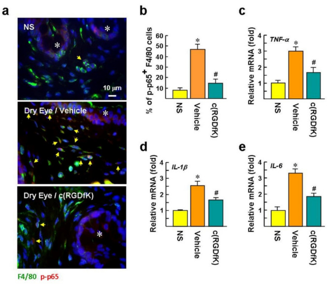
c(RGDfK) suppresses inflammatory responses in dry eyes. The mice received DED induction and c(RGDfK) treatment as described in the legend of Figure 5A. (a,b) Representative images of immunofluorescence staining of NF-κB p-p65 (Ser536) and F4/80 in the conjunctiva after DED induction for 12 days. The p-p65 located in the cell nucleus is confirmed by Hoechst 33258 counterstaining. Asterisks indicate conjunctival epithelium. Arrows indicate p-p65 and F4/80 double-positive cells. There were 30 randomly selected fields in each treatment (n = 6 per group) that were photographed, and the percentage of p-p65 and F4/80 double-positive cells per total F4/80-positive cells was calculated. * p < 0.0006 versus NS mice. # p < 0.0001 versus vehicle group. (c–e) qPCR evaluation of the mRNA levels of proinflammatory cytokines in the conjunctiva after DED induction for 12 days. Values are expressed as mean ± SE. * p < 0.001 versus NS mice. # p < 0.02 versus the vehicle-treated group.

