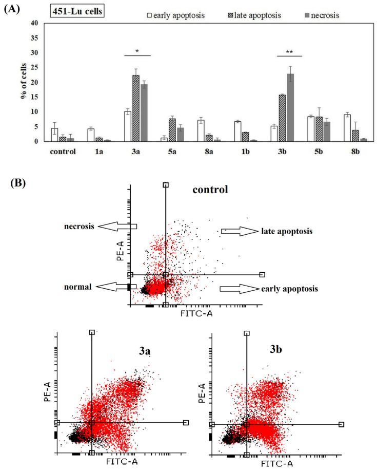
Figure 6.
Induction of apoptosis in 451-Lu cells in the presence of the studied compounds. Cells were cultured for 72 h in the presence of the studied chemicals (at 100 µM). The percentage of necrotic, early and late apoptotic cells was shown. The statistical significance was determined (* p < 0.05 and ** p < 0.001) (A). Representative histograms of events defined after forward and side scatters as cells (red dots) and debris (black dots) are shown (B).

