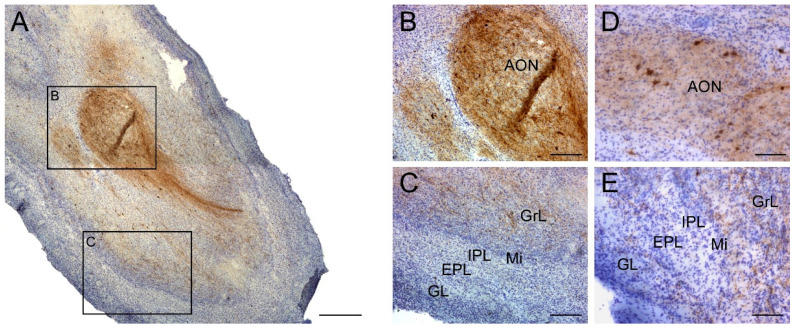
Figure 1.
Immunohistochemistry staining for somatostatin in the olfactory bulb. Somatostatin is present in all regions within the olfactory bulb (A) with a striking presence in the anterior olfactory nucleus (B), and expression was low in the GrL and within the Mi, IPL, EPL and GL external layers in non-AD cases (C). Similarly, SST-cells did not show neurodegeneration features in AD cases and were mainly placed in the anterior olfactory nucleus (D). In addition, lower intensity of labeling in SST -expressing fibers was observable in the rest of olfactory layers in AD cases (E). Note that the number of somatostatin-expressing cells was very low compared with the number of somatostatin fibers. GrL; granule cell layer, Mi; mitral cell layer, IPL; internal plexiform layer, EPL; external plexiform layer, GL; glomerular layer. Scale bar A, 500 µm; B,C, 200 µm.

