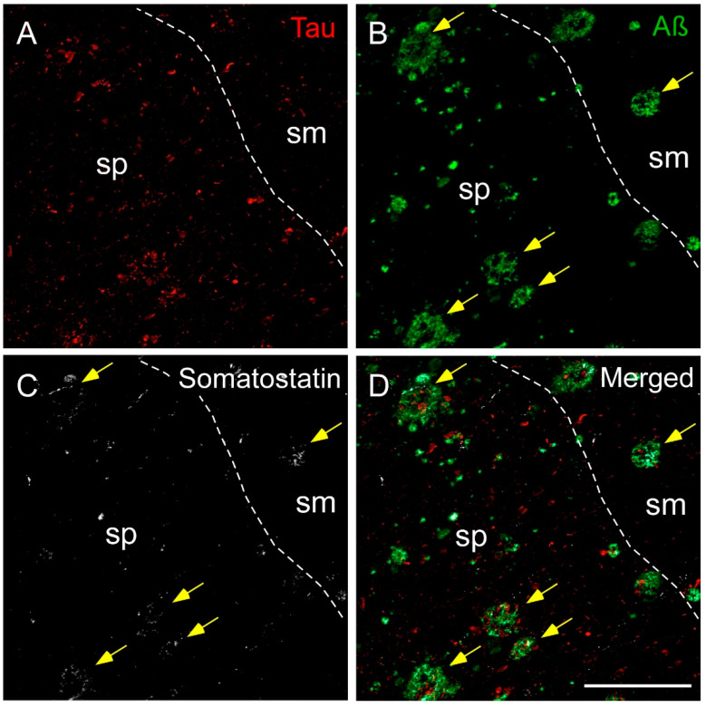
Figure 6.
Immunofluorescence staining of somatostatin, tau and amyloid-β in the human hippocampus in AD. Representative images show staining in the CA1 subfield for tau (A), amyloid-β (B) and somatostatin (C). Tau was mainly located in the pyramidal layer (sp), whereas amyloid plaques localized to both the pyramidal layer and the molecular layer (sm). Somatostatin fibers and cell debris were found in both layers, preferentially coexpressing amyloid-β (yellow arrows) (D). Scale bar A–D 200 µm.

